
What Is Tinea Capitis?
Tinea capitis represents a dermatophyte infection specifically targeting the scalp and hair follicles. Unlike other superficial fungal infections, this condition requires systemic treatment due to its deep penetration into hair shafts and follicular structures.Overview of Scalp Ringworm
Scalp ringworm is a misnomer, as the infection is caused by fungi, not worms. The characteristic circular, scaly patches result from the fungal organism's growth pattern. This infection primarily affects the hair shaft, hair follicle, and surrounding scalp tissue, leading to hair breakage and potential temporary hair loss. The condition occurs worldwide but shows geographic variations in causative organisms. In developed countries, tinea capitis primarily affects children between ages 3-14, with peak incidence in school-age children. The infection spreads rapidly in close-contact environments such as schools, daycare centers, and households.Common Fungi That Cause Tinea Capitis
Several dermatophyte species can cause tinea capitis: Trichophyton tonsurans: Most common in North America, responsible for anthropophilic transmission and typically causes "black dot" tinea capitis with minimal inflammation. Microsporum canis: Common in areas with high pet populations, this zoophilic organism causes gray patch tinea capitis and associates with infected cats and dogs. Microsporum persicolor: Emerging pathogen showing increased prevalence, often causing inflammatory presentations. Trichophyton violaceum: Common in parts of Africa and Asia, causing severe inflammatory reactions with scarring potential.Tinea Capitis Symptoms and Types
Understanding various presentations is crucial for early recognition and appropriate treatment initiation.Common Signs of Scalp Ringworm
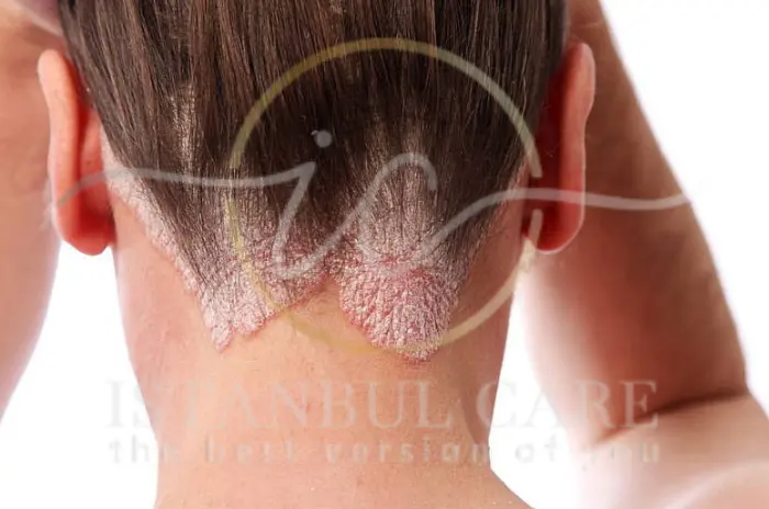
The clinical presentation varies significantly depending on the causative organism, host immune response, and infection duration: Hair Loss Patterns: Patchy areas with irregular borders, often with visible broken hair shafts at different lengths. Affected areas may appear as bald patches or areas with very short, broken hairs. Scaling and Inflammation: Dry, flaky scaling resembling severe dandruff. Scaling may be fine and powdery or thick and adherent, depending on fungal species. Itching and Discomfort: Persistent itching that may worsen at night. Some patients experience tenderness or burning sensations in affected areas. Lymph Node Enlargement: Swollen lymph nodes in the neck region, particularly behind ears, indicating immune response.Black Dot vs. Gray Patch Tinea Capitis
Black Dot Tinea Capitis: Caused primarily by Trichophyton tonsurans, featuring hair breaking at scalp surface leaving dark dots, minimal scaling, subtle presentation, and multiple small patches. Gray Patch Tinea Capitis: Typically caused by Microsporum species, characterized by prominent scaling with gray appearance, hair breaking above scalp surface, obvious visual presentation, and larger defined patches.Inflammatory type with pustules or kerion
Kerion Formation: Most severe inflammatory response presenting as boggy, swollen scalp areas with pus-filled lesions, significant pain, potential permanent scarring, and may be mistaken for bacterial infections. Pustular Variants: Less severe inflammatory responses featuring scattered pustules with increased redness and warmth.How Is Tinea Capitis Diagnosed?
Accurate diagnosis requires clinical assessment and laboratory confirmation for appropriate treatment selection.Clinical Examination and Tools
Visual Assessment: Dermatologists examine scalp for characteristic hair loss patterns, scaling, and inflammation. Distribution and appearance provide clues about causative organisms. Wood's Lamp Examination: Ultraviolet light reveals fluorescence in certain Microsporum infections, though many current strains don't fluoresce. Hair Pull Test: Gentle traction demonstrates ease of hair removal and breakage patterns characteristic of fungal infection.Role of KOH Prep, Cultures, and Dermoscopy
KOH Preparation: Direct microscopic examination of hair samples revealing fungal elements including spores and hyphae, providing rapid preliminary diagnosis within minutes. Fungal Culture: Gold standard requiring 2-4 weeks but providing definitive diagnosis, species identification, and antifungal susceptibility patterns. Dermoscopy: Magnified examination revealing "comma hairs," corkscrew hairs, yellow dots, and black dots providing diagnostic clues.When to Consider a Biopsy or Specialist Referral
Biopsy Indications: Atypical presentations, treatment-resistant cases, suspected secondary infections, or kerion formation with uncertain diagnosis.Specialist Referral: Treatment failure, recurrent infections, immunocompromised patients, or complex multi-family cases.
Causes and Risk Factors of Tinea Capitis
Understanding transmission patterns helps in treatment and prevention strategies.How Do You Get Scalp Fungus?
Direct Human Contact: Person-to-person transmission through direct contact, particularly common in households and schools. Fomite Transmission: Sharing contaminated combs, brushes, hats, helmets, bedding, towels, and salon equipment. Animal Contact: Transmission from infected pets, particularly cats and dogs, plus farm animals and wildlife. Environmental Exposure: Some fungi persist in soil, though less common for scalp infections.Who Is Most at Risk for Tinea Capitis?
Age Factors: Children ages 3-14 show highest susceptibility due to immature immune systems, frequent peer contact, and less developed hygiene practices. Demographic Risks: African American children show higher prevalence, lower socioeconomic status correlates with increased risk, and overcrowded conditions facilitate transmission. Medical Risks: Immunocompromised states, chronic skin conditions, previous fungal infections, and certain genetic factors influence susceptibility.Tinea Capitis Treatment Options
Effective treatment requires systemic antifungal medications, as topical treatments alone are insufficient.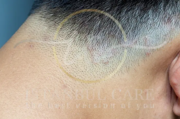
First-Line Antifungal Medications
Systemic Therapy Requirements: Oral medications are necessary because topical agents cannot penetrate hair follicles adequately, and infection involves internal hair shaft structures.
Primary Options: Griseofulvin (traditional first-line), terbinafine (newer agent), itraconazole (resistant cases), and fluconazole (specific regions/resistance).
Griseofulvin vs. Terbinafine
| Medication | Advantages | Disadvantages | Best For |
|---|---|---|---|
| Griseofulvin | Extensive safety data, liquid available | Longer duration, interactions | Microsporum, young children |
| Terbinafine | Shorter course, fewer interactions | Limited pediatric data | Trichophyton, older children |
Griseofulvin: Requires 6-12 weeks, better absorption with fatty foods, established pediatric safety, more effective against Microsporum.
Terbinafine: Typically 4-6 weeks, superior against Trichophyton tonsurans, fewer interactions, better compliance.
Duration and Dosage Recommendations
Griseofulvin Dosing: Microsize 20-25 mg/kg/day (max 1000mg), ultramicrosize 15-20 mg/kg/day (max 750mg), continue 2-4 weeks after clinical resolution.
Terbinafine Dosing: 10-20 kg (62.5 mg daily), 20-40 kg (125 mg daily), >40 kg (250 mg daily), 4-6 weeks duration.
Adjunctive Topical Therapies
Selenium Sulfide Shampoo: Reduces surface spore burden, decreases transmission risk, use 2-3 times weekly during treatment.
Ketoconazole Shampoo: Alternative with antifungal properties, may reduce treatment duration when combined with oral therapy.
Preventing Tinea Capitis
Prevention focuses on reducing transmission risk and environmental contamination.
Tips to Avoid Scalp Fungal Infections
Personal Hygiene: Regular hair washing with antifungal shampoos in high-risk individuals, avoid sharing personal items, maintain good hygiene, prompt treatment of suspicious symptoms.
Environmental Precautions: Regular cleaning of shared spaces, proper pet care, awareness of household symptoms, education about transmission risks.
Cleaning and Disinfecting to Stop Spread
Household Protocol: Clean accessories with 10% bleach solution, wash bedding in hot water (>140°F), vacuum thoroughly, disinfect surfaces.
Personal Items: Replace or disinfect brushes and combs, avoid sharing during treatment, consider disposable accessories, clean salon tools between uses.
Managing Contagion and Prevention
Understanding contagion patterns helps implement appropriate measures.
Is Tinea Capitis Contagious?
Highly contagious until effective treatment begins showing results (typically 2-4 weeks). Contagion decreases gradually: pre-treatment (highly contagious), early treatment (decreasing), mid-treatment (significantly reduced), late treatment (minimal risk).
Hygiene and Household Measures
Family Screening: Examine all household members, particularly children and close contacts.
Isolation Recommendations: Avoid head-to-head contact, separate beds during early treatment, use separate towels, implement strict hand hygiene.
School and Daycare Guidelines
Return Policies: Most institutions allow return after 2-4 weeks of treatment with clinical improvement.
Notification: Many schools require notification for preventive measures and contact tracing.
Complications and Prognosis
Understanding potential complications guides management decisions.
Potential for Hair Loss or Scarring
Temporary Hair Loss: Most common outcome with complete regrowth within 6-12 months. Hair typically returns to normal texture and color.
Permanent Scarring: Rare but possible with delayed treatment, severe inflammation, secondary infections, or immunocompromised status.
Risk of Kerion and Secondary Infections
Kerion Development: Severe inflammatory response from delayed treatment, inappropriate corticosteroids, zoophilic organisms, or bacterial superinfection.
Management: May require additional antibiotics and specialized wound care.
Aftercare and Follow-Up
Proper follow-up ensures treatment success and prevents recurrence.
What to Expect After Treatment Starts
Timeline: Week 1-2 (reduced itching/scaling), Week 2-4 (decreased inflammation/new growth), Week 4-8 (significant improvement), Month 2-6 (complete regrowth).
Monitoring: Regular assessment of response, side effects, and compliance.
How to Reduce the Risk of Reinfection
Environmental Decontamination: Thorough cleaning of contaminated items and surfaces.
Family Treatment: Screen and treat all affected household members simultaneously.
Ongoing Vigilance: Regular scalp examination for early symptom detection.
What to Expect After Treatment
Duration of Treatment and Follow‑Up
Monitoring Schedule: Week 2 (early response), Week 4-6 (improvement evaluation), Week 8-12 (completion assessment), Month 3-6 (resolution follow-up).
Laboratory Monitoring: Periodic liver function tests for prolonged therapy patients.
Avoiding Permanent Hair Loss After Recovery
Gentle Care: Use mild shampoos, avoid aggressive brushing during recovery.
Nutritional Support: Ensure adequate protein, iron, and vitamins for healthy regrowth.
Patience: Complete restoration typically takes 6-12 months after successful treatment.
We’re ready to answer your questions
Tinea capitis is a fungal infection of the scalp and hair follicles caused by dermatophyte organisms. Also known as scalp ringworm, it primarily affects children and causes patchy hair loss, scaling, and inflammation. The infection penetrates deep into hair shafts and follicles, requiring systemic antifungal treatment for effective cure.
Common symptoms include patchy hair loss with irregular borders, dry flaky scaling, persistent itching, broken hair shafts at various lengths, and swollen neck lymph nodes. Appearance varies by species - some cause "black dot" patterns with hair breaking at scalp level, others create "gray patch" patterns with obvious scaling.
Diagnosis involves clinical examination followed by laboratory testing. Doctors use KOH preparation for immediate microscopic examination, fungal cultures for definitive identification (2-4 weeks), and dermoscopy to identify characteristic features. Wood's lamp may reveal fluorescence in certain infections.
Spreads through direct contact with infected people, sharing contaminated items like combs and hats, contact with infected animals, and rarely environmental exposure. Children ages 3-14 are most at risk due to immature immunity, frequent peer contact, and developing hygiene practices.
Main treatments are oral antifungals including griseofulvin (6-12 weeks, best for Microsporum) and terbinafine (4-6 weeks, superior for Trichophyton). Other options include itraconazole and fluconazole for resistant cases. Topical treatments alone are insufficient.
Highly contagious until effective treatment shows results (2-4 weeks). Prevention involves avoiding sharing personal items, maintaining hygiene, cleaning with bleach solution, washing bedding in hot water, and screening contacts. Schools typically allow return after 2-4 weeks of treatment with improvement.
Most cases result in temporary hair loss with complete regrowth within 6-12 months. Permanent scarring is rare but possible with delayed treatment, severe inflammation, or secondary infections. Early treatment significantly improves outcomes and reduces scarring risk.
Treatment lasts 4-12 weeks depending on medication. Expect reduced itching within 1-2 weeks, new hair growth by 2-4 weeks, and significant improvement by 4-8 weeks. Complete regrowth takes 6-12 months. Regular follow-up monitors progress, and professionals at our intermediary organization provide ongoing support throughout treatment.
Follow us on social media for updates, tips, and patient success stories: